Colour Doppler Study in Lucknow
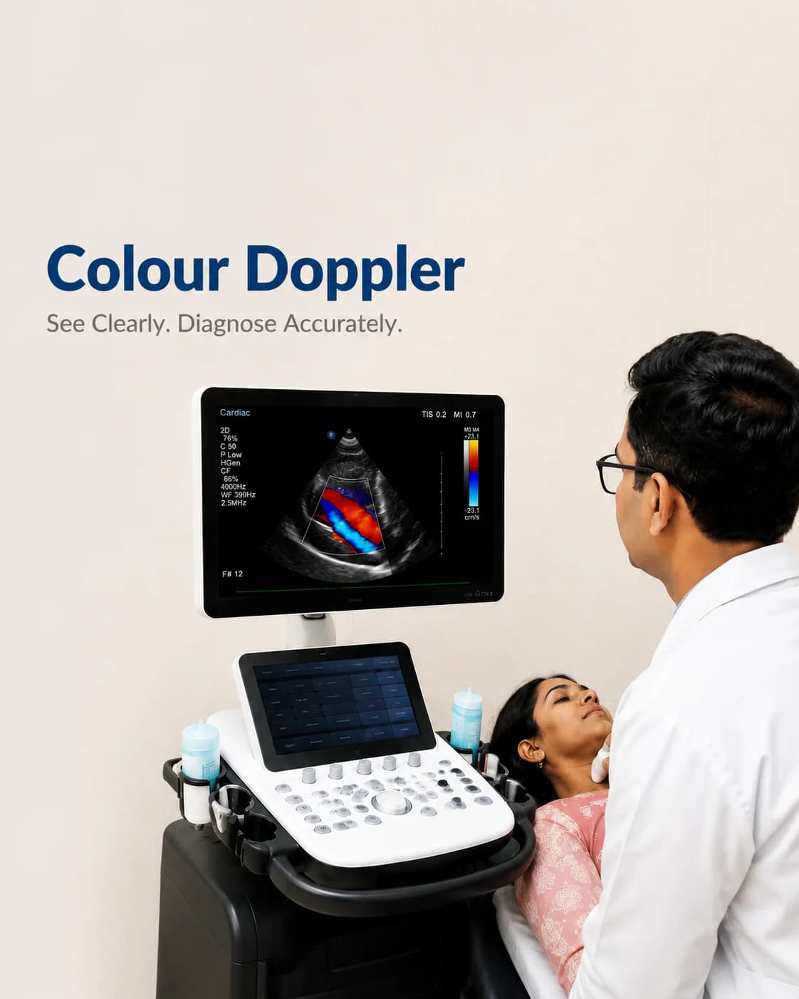
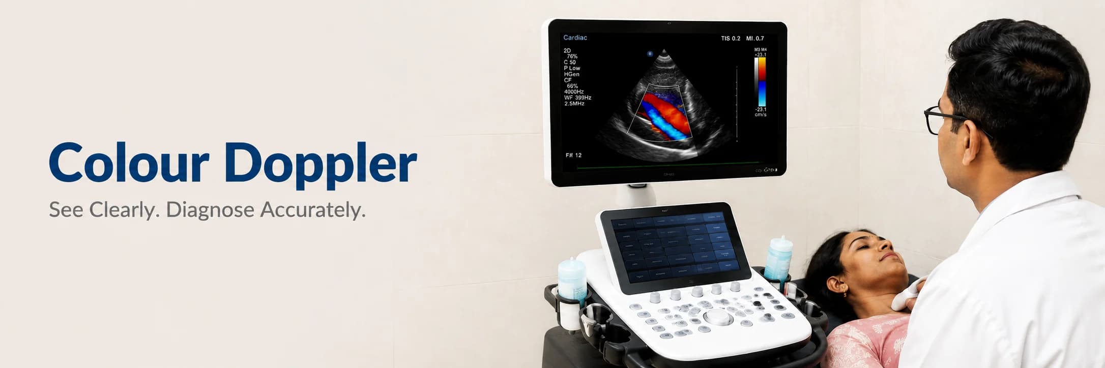
Ultrasound that maps blood flow in colour — through the heart, arteries and veins.
A colour Doppler study is an ultrasound that shows not just the structure of a blood vessel but the blood moving through it, mapped in colour. It reveals a narrowed artery, a clot in a vein, or blood flowing the wrong way through a valve — all without radiation and without any discomfort.
At Charak, Doppler studies of the heart, the neck arteries, and the arteries and veins of the limbs are performed by trained operators and reported by specialists. It turns a still picture of a vessel into a study of how well blood is actually getting through it.
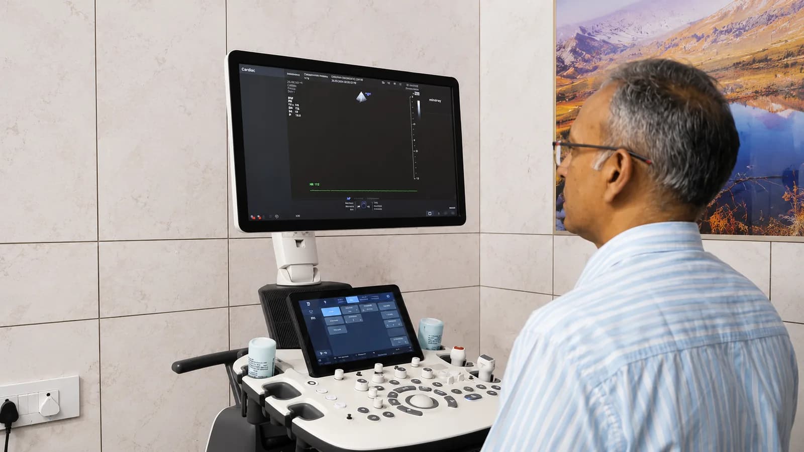
What colour Doppler reveals
By colouring blood flow — typically one colour towards the probe and another away from it — Doppler shows the direction and speed of flow. A tight narrowing speeds the blood up; a blockage stops it; a leaking valve sends it backwards. Each has a signature the study can measure.
This is how a carotid Doppler assesses stroke risk from narrowed neck arteries, how a venous Doppler finds a deep vein clot in a swollen leg, and how cardiac Doppler grades a valve problem.
A colour Doppler study may be recommended for
- Leg swelling or pain, to look for a deep vein clot
- Assessing narrowing in the neck (carotid) arteries and stroke risk
- Poor circulation, cramps or non-healing wounds in the limbs
- Grading a heart valve problem (cardiac Doppler)
- Checking blood flow to an organ or a graft
How to prepare
Preparation depends on which vessels are being studied — most need none. We tell you what yours needs when you book.
- Neck (carotid), limb and cardiac Doppler: no special preparation, no fasting
- Some abdominal vessel studies need a few hours' fasting — we will tell you if yours does
- Wear clothing that lets the area being scanned be reached easily
- Bring any previous Doppler or vascular reports for comparison
What to expect
- 01
Settling in
You lie or sit so the area to be studied is accessible, and gel is applied for the probe.
- 02
The scan
The operator moves the probe over the vessels, watching the flow in colour and measuring its speed.
- 03
Listening to flow
You may hear a rhythmic, whooshing sound — that is the blood flow being measured, and it is entirely normal.
- 04
Afterwards
The gel is wiped off and you can leave immediately; the specialist's report follows.
Common questions
Does a Doppler study hurt?
No. It is an ultrasound — you feel only the gentle pressure of the probe and the cool gel. Some studies press a little more firmly to find a deep vessel, but it is not painful.
What is the colour on the screen showing?
The colour maps blood flow — usually one colour for flow towards the probe and another for flow away from it. It lets the operator see the direction and speed of blood, and spot a narrowing, a clot, or a leaking valve.
Why would I need a carotid Doppler?
It checks the arteries in the neck that supply the brain for narrowing, which is a cause of stroke. Finding and grading that narrowing helps your doctor reduce the risk before a stroke happens.
From the health library
Plain-language guides written by the team here — what the test measures, how to prepare, and what the result means.