
MRI Scan in Lucknow — Magnetic Resonance Imaging
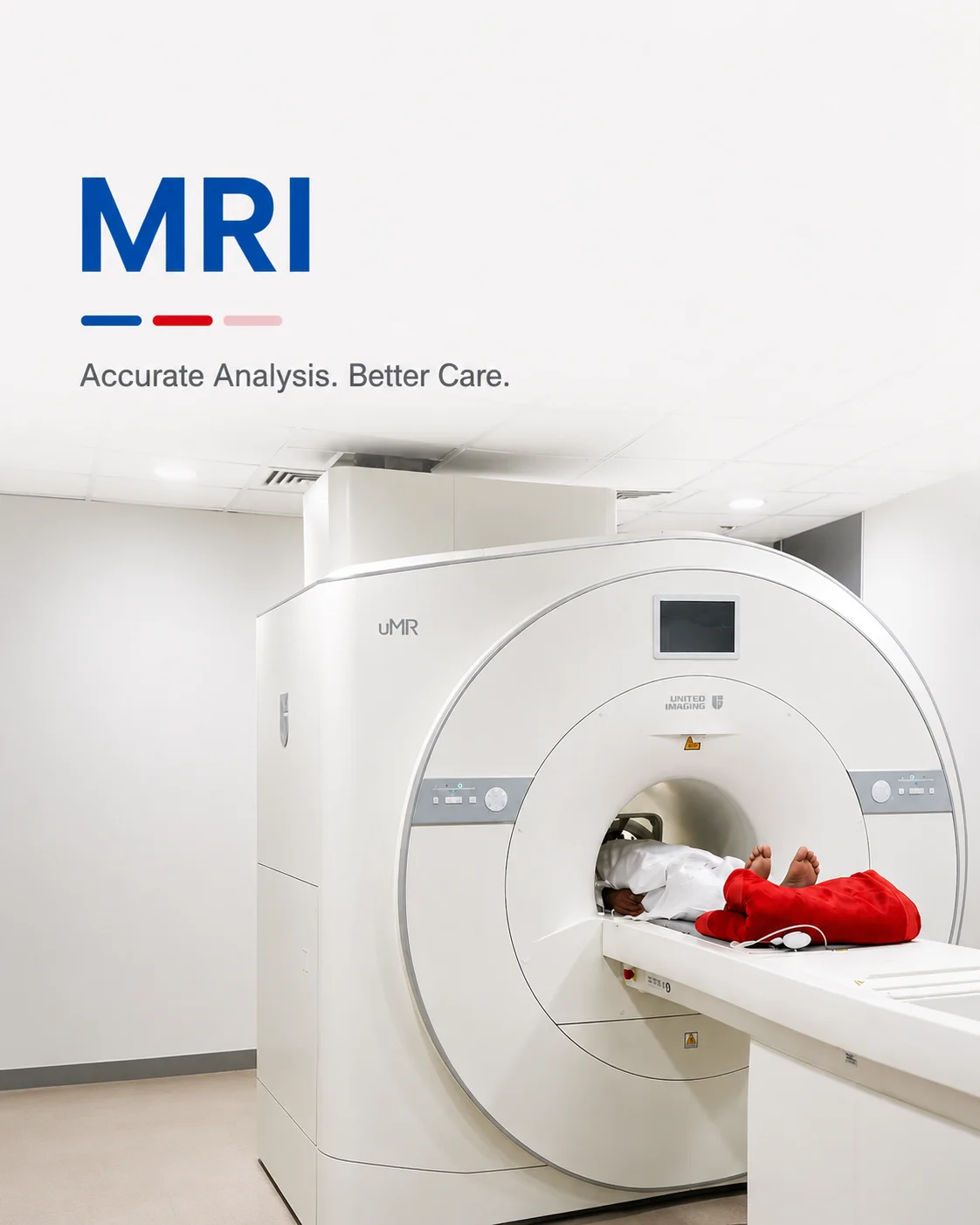
Detailed, radiation-free imaging of the brain, spine, joints and soft tissue.
Magnetic Resonance Imaging (MRI) uses a strong magnetic field and radio waves — not X-rays — to build highly detailed pictures of the soft tissues inside your body. It is the imaging of choice when a doctor needs to see the brain, spinal cord, ligaments, cartilage or abdominal organs clearly, and it involves no ionising radiation at all.
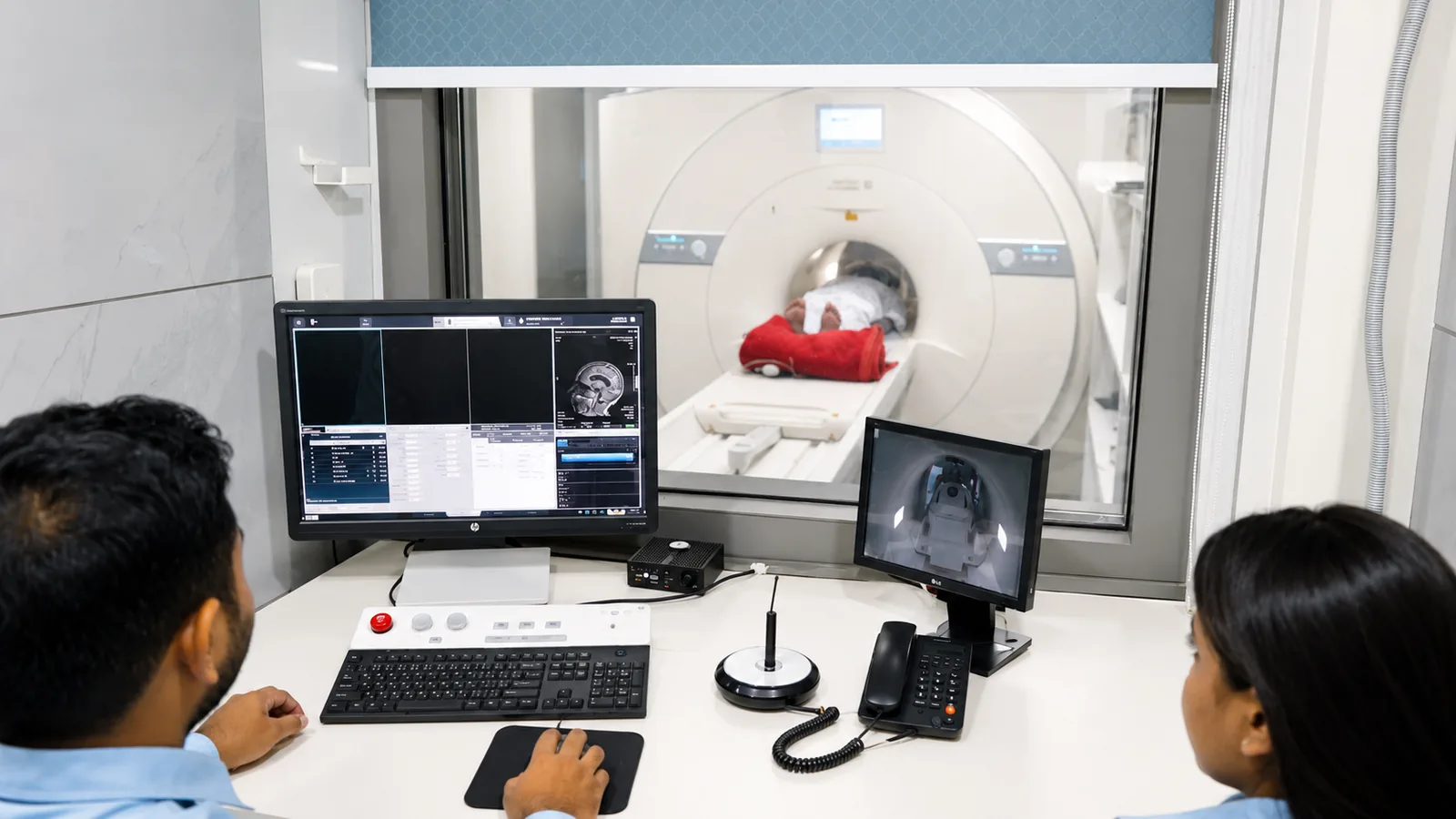
At Charak, MRI studies are acquired on high-field equipment and reported by consultant radiologists, so the scan and its interpretation are held to the same standard. Where a previous study exists, it is compared rather than read in isolation — a change over time is often the finding that matters most.
What an MRI shows, and when it is the right scan
Because MRI distinguishes between soft tissues so well, it excels at questions that X-ray and CT cannot fully answer: the detail of a slipped disc pressing on a nerve, an early stroke, a torn knee ligament, or the internal structure of the liver and pelvis.
It is also the safe choice when imaging needs to be repeated over months or years, because there is no cumulative radiation dose to weigh against the benefit — a real advantage in following a known condition.
Your doctor may recommend an MRI for
- Persistent headache, seizures, or suspected stroke (brain MRI)
- Back or neck pain with numbness, weakness or sciatica (spine MRI)
- Knee, shoulder or other joint injury — ligaments, cartilage, tendons
- Abdominal or pelvic conditions needing soft-tissue detail
- Follow-up of a known tumour, lesion or inflammatory condition
How to prepare
The magnetic field is always on. Tell us before your appointment about any pacemaker, cochlear implant, metal fragment, or surgical implant — this screening is the single most important safety step.
- Wear comfortable clothing with no metal — zips, hooks or embroidery with metallic thread
- Leave jewellery, watches and hairpins at home or hand them in before the scan
- For most MRIs you can eat and drink normally; some abdominal studies need a short fast, and we will tell you if so
- If you know you are claustrophobic, tell us in advance — we can plan for it
- Bring any previous scans or films so the radiologist can compare
What to expect on the day
- 01
Safety screening
A radiographer checks the safety questionnaire with you and confirms there is nothing that the magnetic field could affect.
- 02
Positioning
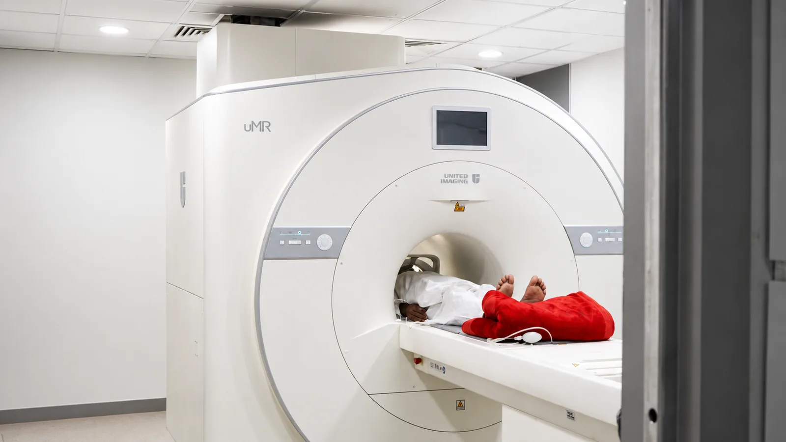
You lie on a padded table that moves into the scanner. You will be given headphones or earplugs — the scan is loud, and that is normal.
- 03
The scan
Each sequence lasts a few minutes; staying still is what keeps the images sharp. Most studies take 20–45 minutes depending on the region and whether contrast is used.
- 04
Contrast, if needed
Some studies use a gadolinium contrast injection to highlight blood vessels or lesions. You will be told beforehand if yours does.
Common questions
Is an MRI safe? Does it use radiation?
MRI uses a magnetic field and radio waves, not ionising radiation, so there is no radiation dose. The main safety concern is metal, which is why implant and pacemaker screening is done carefully before every scan.
How long does an MRI take?
Most scans take between 20 and 45 minutes, depending on the body part and whether contrast is used. You will be able to hear from and speak to the radiographer throughout.
Will I feel closed in?
The scanner is open at both ends and well lit and ventilated. If you are prone to claustrophobia, tell us in advance so we can plan the scan to keep you comfortable.
Can I eat before my MRI?
For most MRIs, yes — eat and take your medicines as usual. A few abdominal studies need a short fast, and we will tell you clearly if yours is one of them.
From the health library
Plain-language guides written by the team here — what the test measures, how to prepare, and what the result means.
Related services
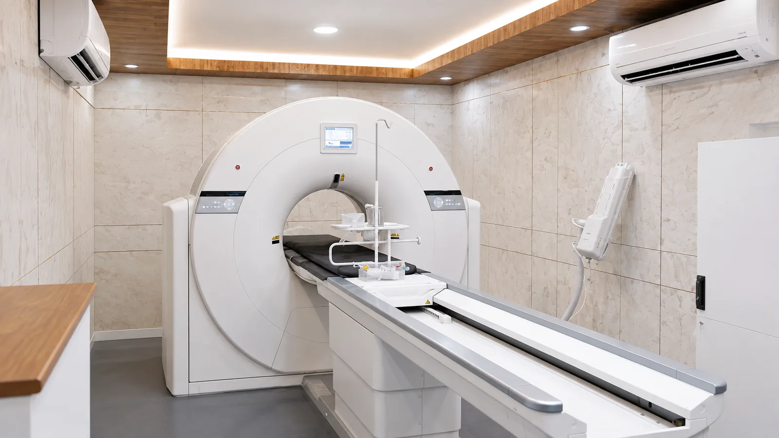
CT Scan
Fast, high-resolution cross-sections of the head, chest, abdomen and blood vessels.
Read more →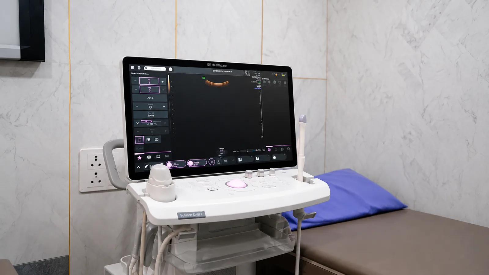
Ultrasound & Doppler
Safe, radiation-free imaging of the abdomen, pelvis, pregnancy and blood flow — in real time.
Read more →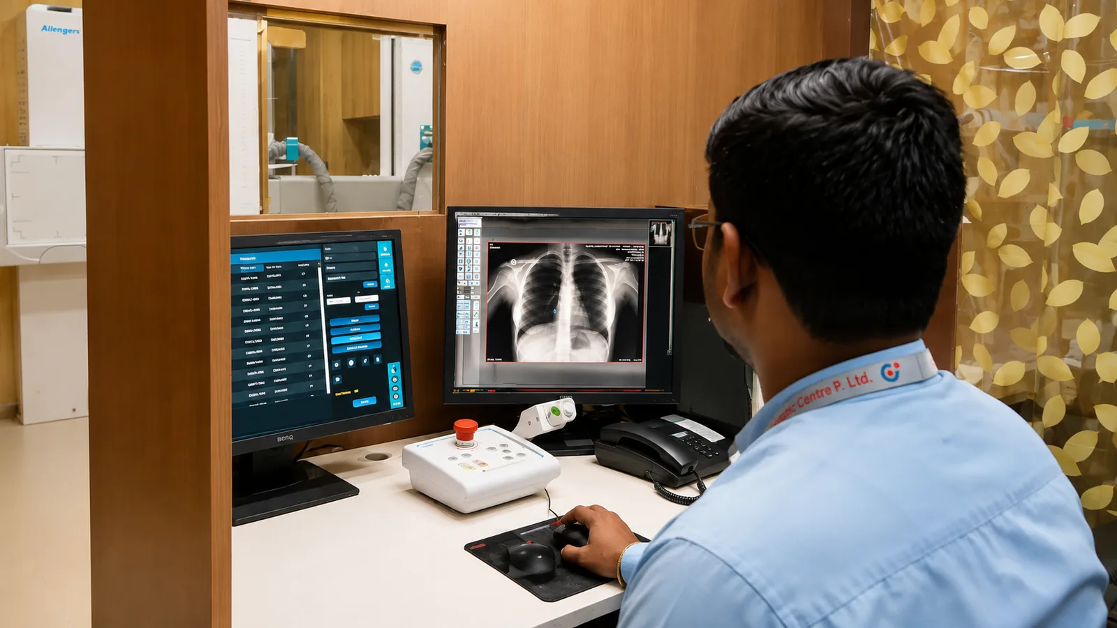
Digital X-Ray
Fast, low-dose imaging of the chest, spine, abdomen and bones — with no film to wait for.
Read more →