
Mammography in Lucknow — Breast Screening & Diagnosis
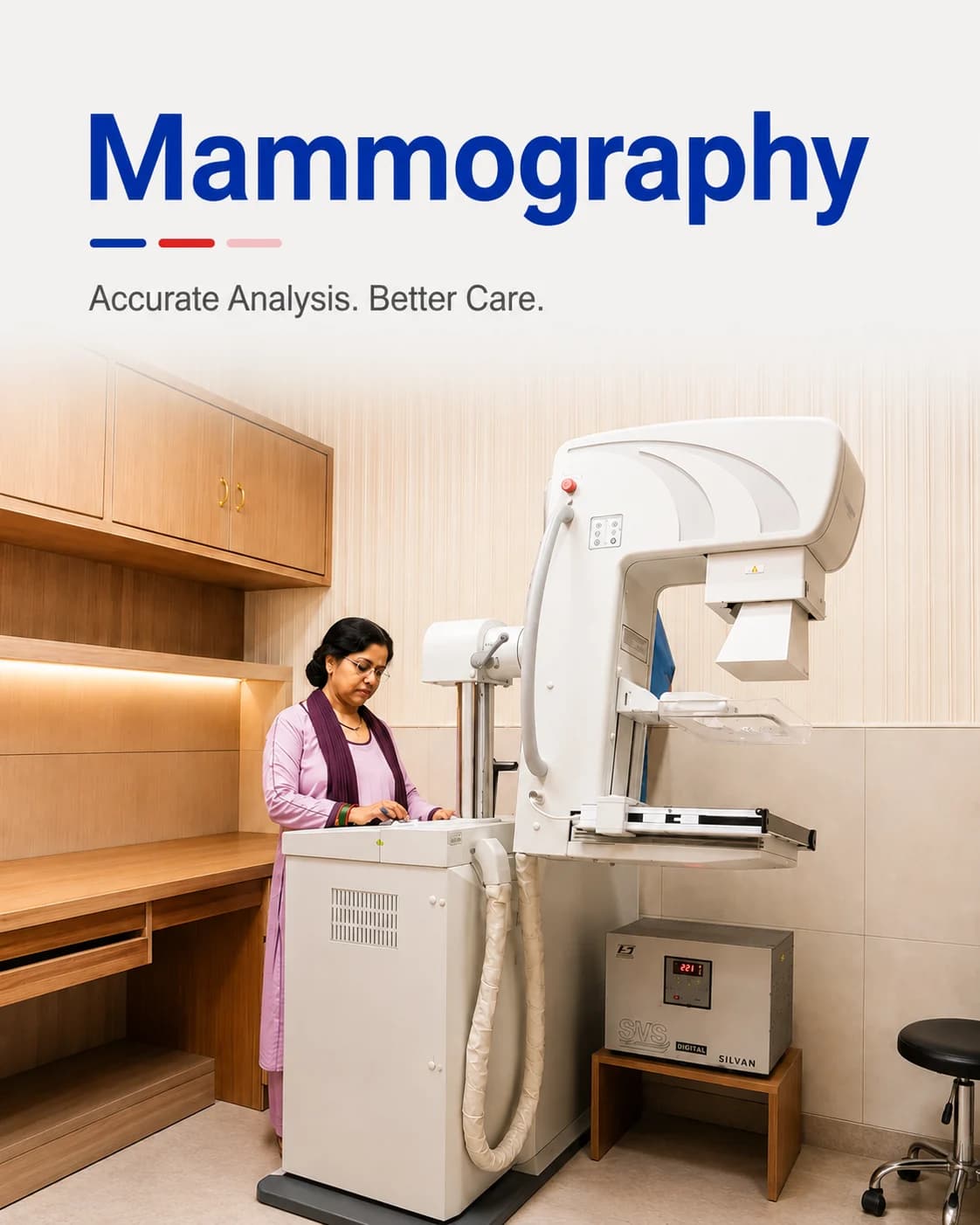
Low-dose breast imaging for screening and for looking into a specific finding.
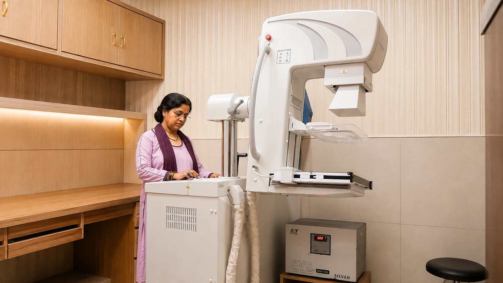
Mammography is a low-dose X-ray of the breast. As a screening test it can find changes long before they can be felt, and as a diagnostic test it looks more closely into a lump or a symptom. Detecting a change early is what gives treatment its best chance.
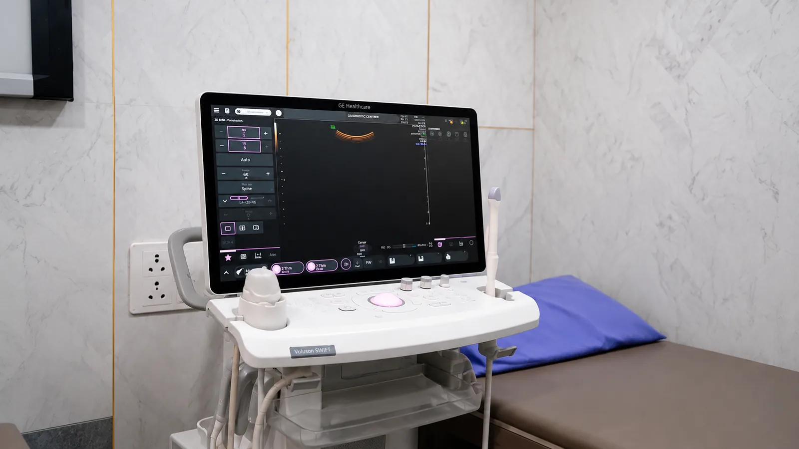
At Charak, examinations are scheduled and staffed so that privacy and dignity are preserved throughout. Where breast density or a specific finding makes it appropriate, mammography is combined with ultrasound so the two together give a fuller answer — and findings that need onward referral are explained, not just printed.
Screening versus diagnostic mammography
A screening mammogram is done when there are no symptoms, to look for early change — usually from the age your doctor advises, and repeated at intervals.
A diagnostic mammogram is done when there is a specific concern — a lump, pain, or a change in the skin or nipple — and takes additional, focused views. Ultrasound is often added, particularly in denser breast tissue where it sees what a mammogram alone may not.
Mammography may be recommended for
- Routine breast screening from the age your doctor advises
- A new breast lump or thickening
- Breast pain that persists or is localised
- A change in the skin, shape or nipple
- Follow-up after previous breast treatment
How to prepare
If you have breast implants or have had breast surgery, tell us when you book so we can plan the right views.
- Book for the week after your period if you still menstruate — the breasts are less tender then
- Do not use talcum powder, deodorant or lotion on the day, as these can show up on the image
- Wear a two-piece outfit so only the upper half needs to be removed
- Bring any previous mammograms for comparison — change over time is important
What to expect on the day
- 01
In private
The examination is done in a private room by a female radiographer, and you undress only from the waist up.
- 02
The views
Each breast is rested on the platform and gently compressed for a few seconds while the image is taken. The pressure is brief and is what gives a clear, low-dose image.
- 03
Ultrasound, if added
If ultrasound correlation is appropriate, it follows immediately in the same visit and is painless.
- 04
Afterwards and reporting
You can dress and leave straight away. A consultant radiologist reports the study, and anything needing follow-up is explained to you clearly.
Common questions
Is a mammogram painful?
The breast is compressed for a few seconds for each view, which can feel uncomfortable but is brief. Booking for the week after your period, when the breasts are less tender, makes it easier.
At what age should I start screening?
It depends on your personal and family history, so your doctor is the best guide. Mammography can begin earlier where there is a family history of breast cancer.
Why is ultrasound sometimes added?
In denser breast tissue, ultrasound can show detail a mammogram alone may miss. Using both together gives a more complete answer, which is why they are often combined.
From the health library
Plain-language guides written by the team here — what the test measures, how to prepare, and what the result means.
 Tumour markers: why they are a poor cancer screening testSold as cancer screening in many packages, and genuinely unsuited to it. What these tests are for, and what actually screens for cancer.6 min read
 Mammography in Lucknow: when to start, and what the discomfort is forThe test people postpone most, for a reason that lasts a few seconds. What it finds, when to start, and how to make it easier.5 min read
Related services
Ultrasound & Doppler
Safe, radiation-free imaging of the abdomen, pelvis, pregnancy and blood flow — in real time.
Read more →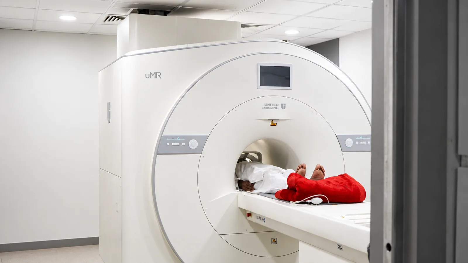
MRI
Detailed, radiation-free imaging of the brain, spine, joints and soft tissue.
Read more →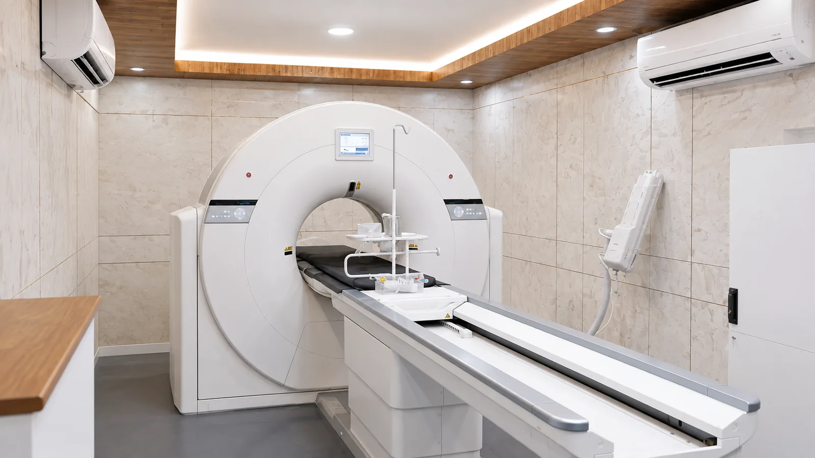
CT Scan
Fast, high-resolution cross-sections of the head, chest, abdomen and blood vessels.
Read more →