
Bone Scan in Lucknow — Nuclear Bone Imaging
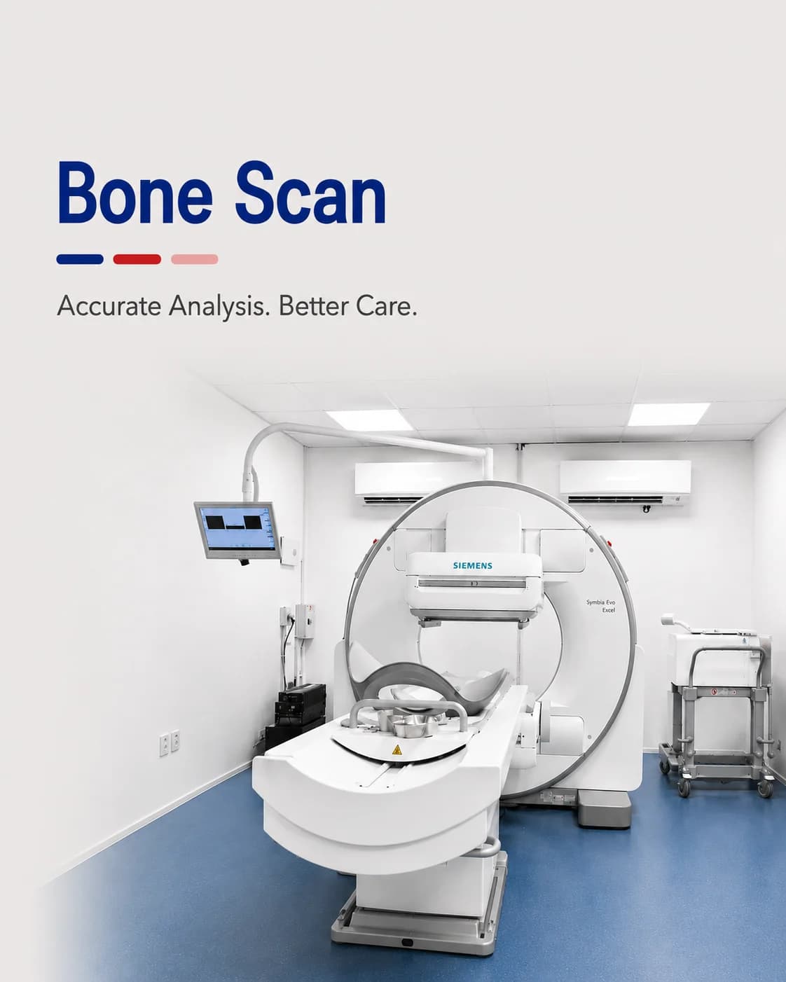
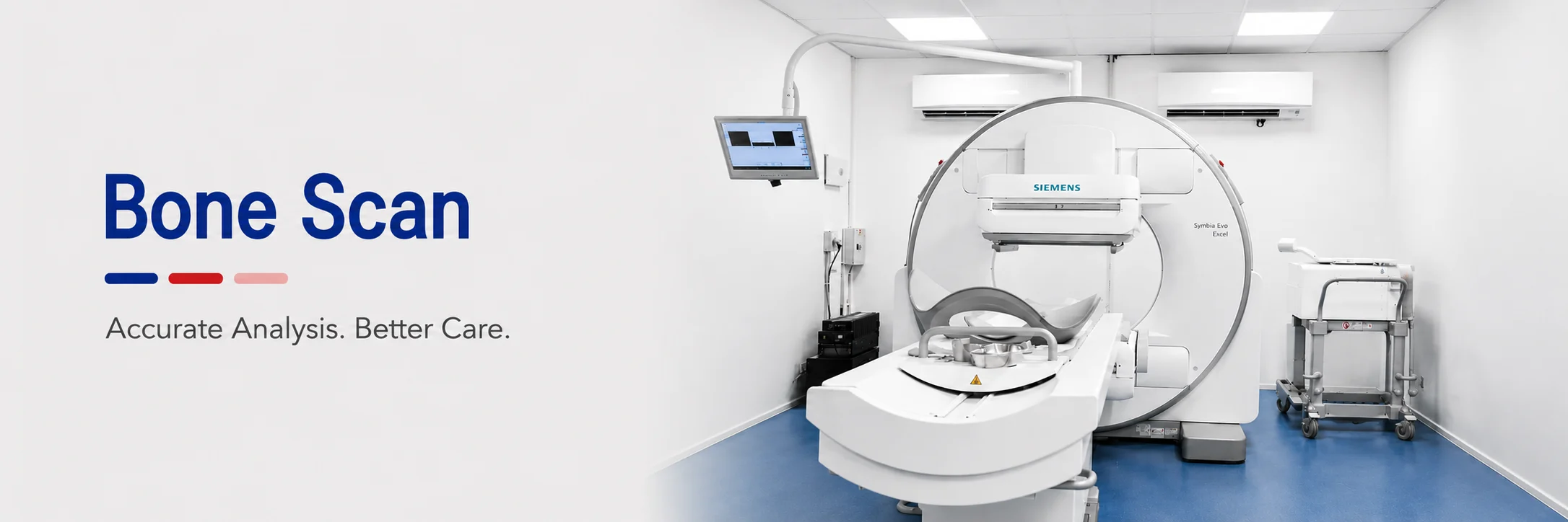
A whole-skeleton study that finds active bone disease before an X-ray can.
A bone scan images the whole skeleton at once, showing where bone is actively repairing or reacting. Because that activity rises long before a change is visible on an X-ray, a bone scan can find a problem — a cancer spread, an infection, a subtle fracture — weeks earlier.
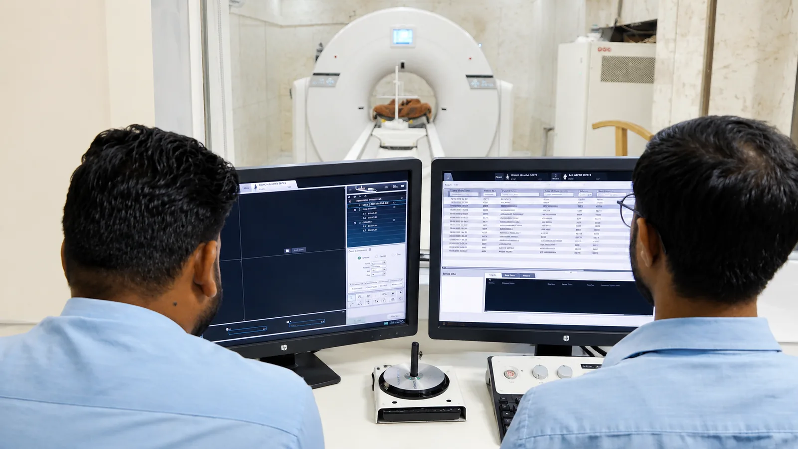
At Charak, bone scans use a tracer that concentrates wherever bone metabolism is high, and are reported by nuclear medicine physicians. One study covers the entire skeleton, which is why it is the test of choice when disease could be anywhere in the bones.
Why a bone scan sees what an X-ray cannot
An X-ray shows a fracture or a lesion once it has changed the bone's structure. A bone scan shows the biological activity around it — the repair, the inflammation — which begins much earlier and shows up as a bright spot on the scan.
It is also comprehensive: rather than X-raying one area at a time, a single bone scan surveys the whole skeleton, which is essential when looking for the spread of a cancer or an unexplained pain that could be anywhere.
A bone scan may be recommended for
- Looking for the spread of cancer to the bones
- Unexplained bone pain that X-rays cannot account for
- A suspected stress fracture not seen on X-ray
- A possible bone infection (osteomyelitis)
- Assessing certain arthritis and metabolic bone conditions
How to prepare
A bone scan has a built-in wait: the tracer is given first, and the scan follows a few hours later once it has settled into the bone. Plan for a return visit or a wait on the day.
- After the tracer injection, drink plenty of water and pass urine often — it clears the tracer from soft tissue for a clearer image
- There is usually a 2–4 hour gap between the injection and the scan
- Remove metal objects before imaging; you may be asked to empty your bladder just before
- Tell us if you are, or might be, pregnant, or are breastfeeding
What to expect on the day
- 01
Tracer injection
The tracer is given through a vein in the arm; the injection itself is quick.
- 02
The uptake wait
You wait a few hours, drinking water, while the tracer is taken up by the bone. You can usually leave and return.
- 03
The scan
You lie still while the gamma camera passes slowly over the whole body — this part takes 20–40 minutes.
- 04
Afterwards
You can resume normal activity; continuing to drink water clears the remaining tracer over the day.
Common questions
Why is there a wait between the injection and the scan?
The tracer needs a few hours to be taken up by the bone and to clear from the surrounding soft tissue. Scanning too early gives a blurred picture, so the delay is what makes the images clear.
Why can a bone scan find things before an X-ray?
An X-ray only shows a problem once it has changed the bone's structure. A bone scan shows the biological activity — repair and inflammation — that begins earlier, so it can detect disease sooner.
Is a bone scan safe?
Yes. The tracer dose is small and short-lived, and drinking water afterwards helps clear it quickly. The information it gives about the whole skeleton is well worth the minimal exposure.
From the health library
Plain-language guides written by the team here — what the test measures, how to prepare, and what the result means.
Related services
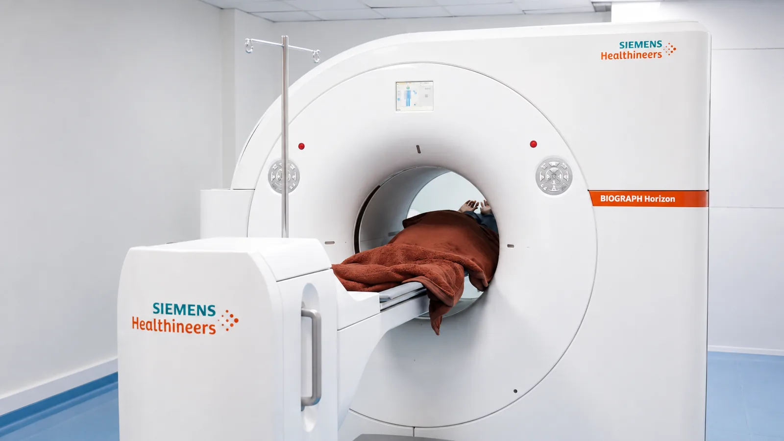
PET-CT Scan
Whole-body imaging that shows how tissue is functioning, not just how it looks.
Read more →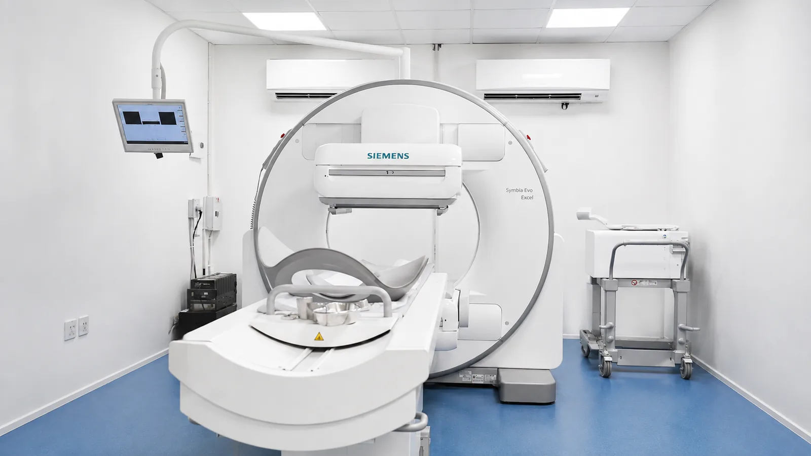
Gamma Camera / SPECT
Functional imaging of the heart, bones, kidneys and thyroid using a gamma camera.
Read more →
Renal Scan (DTPA / DMSA)
Measuring how well each kidney drains and works — one side against the other.
Read more →