Ultrasound & Colour Doppler in Lucknow
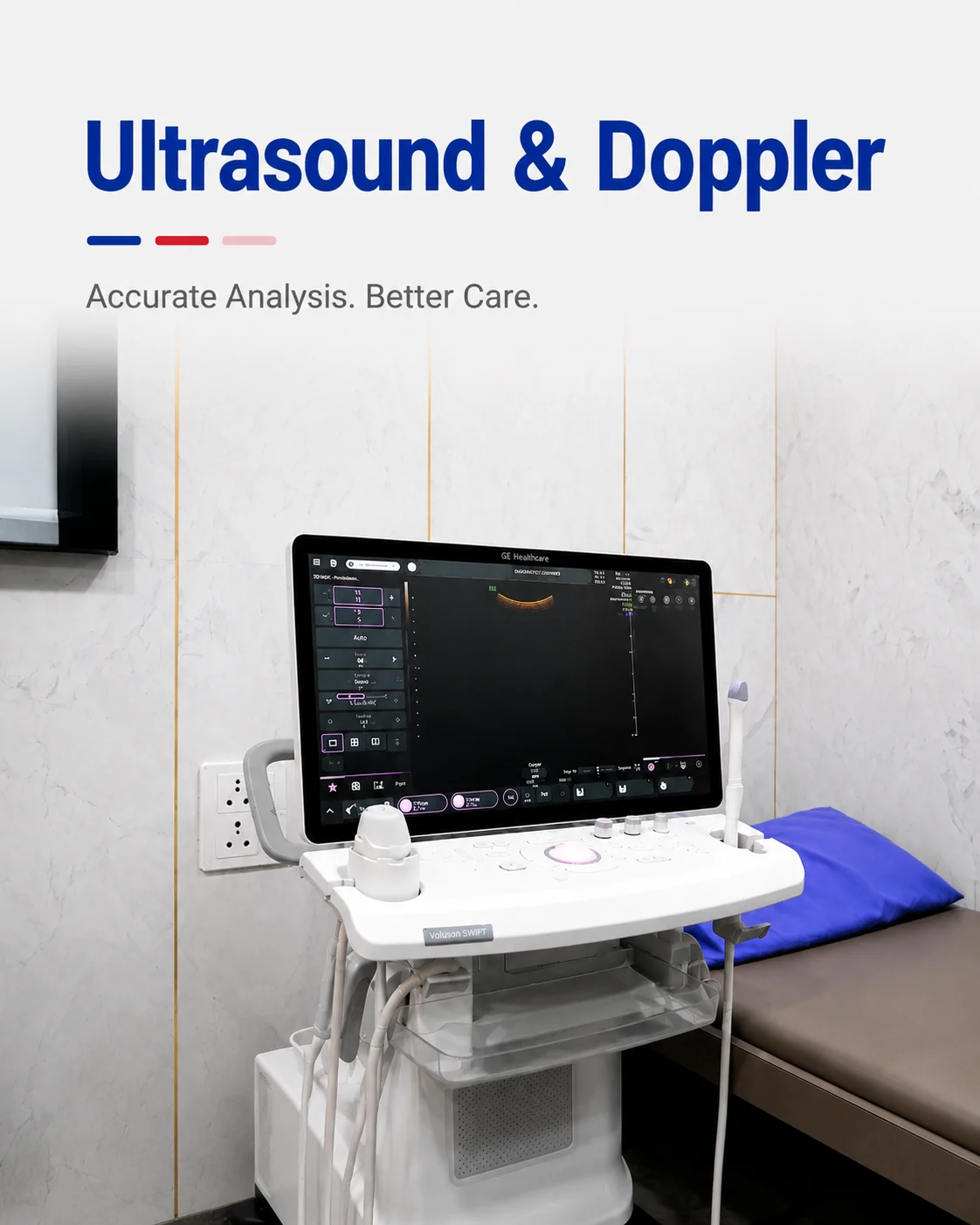
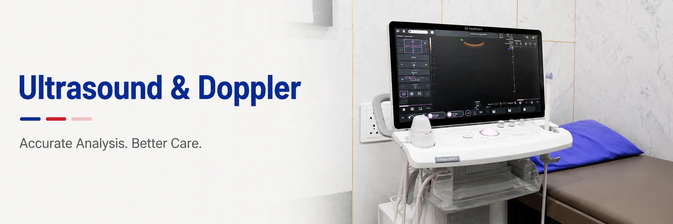
Safe, radiation-free imaging of the abdomen, pelvis, pregnancy and blood flow — in real time.
Ultrasound uses high-frequency sound waves — no radiation — to produce moving, real-time images of the organs, blood vessels and, in pregnancy, the baby. It is completely safe to repeat, which is why it is used throughout pregnancy and to follow many conditions over time.
Ultrasound is more operator-dependent than any other scan: the quality of the study is decided at the probe, not at the report. At Charak, scans are performed by trained sonographers and radiologists rather than delegated, so the images the report is based on are reliable.
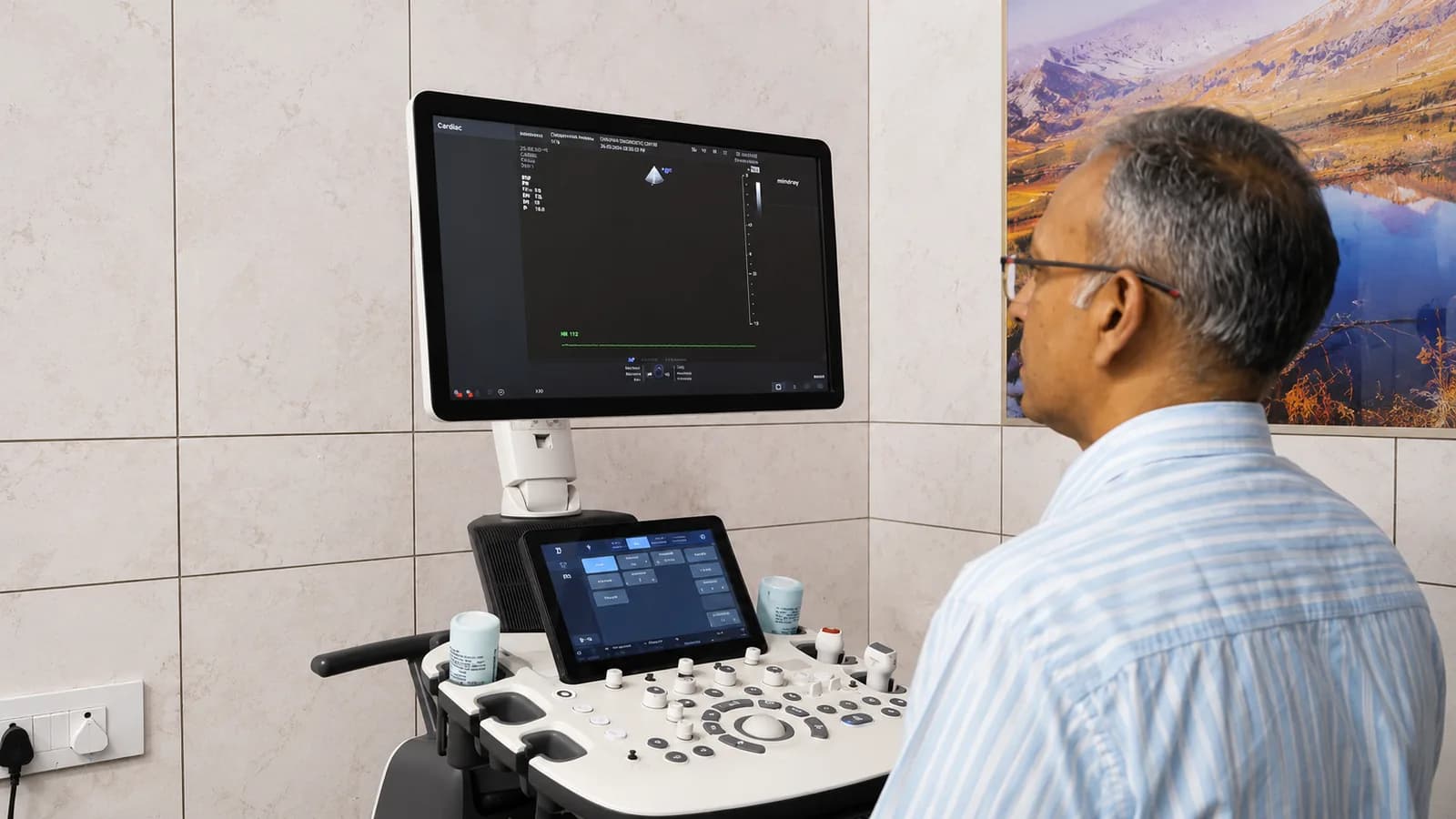
What ultrasound and Doppler can see
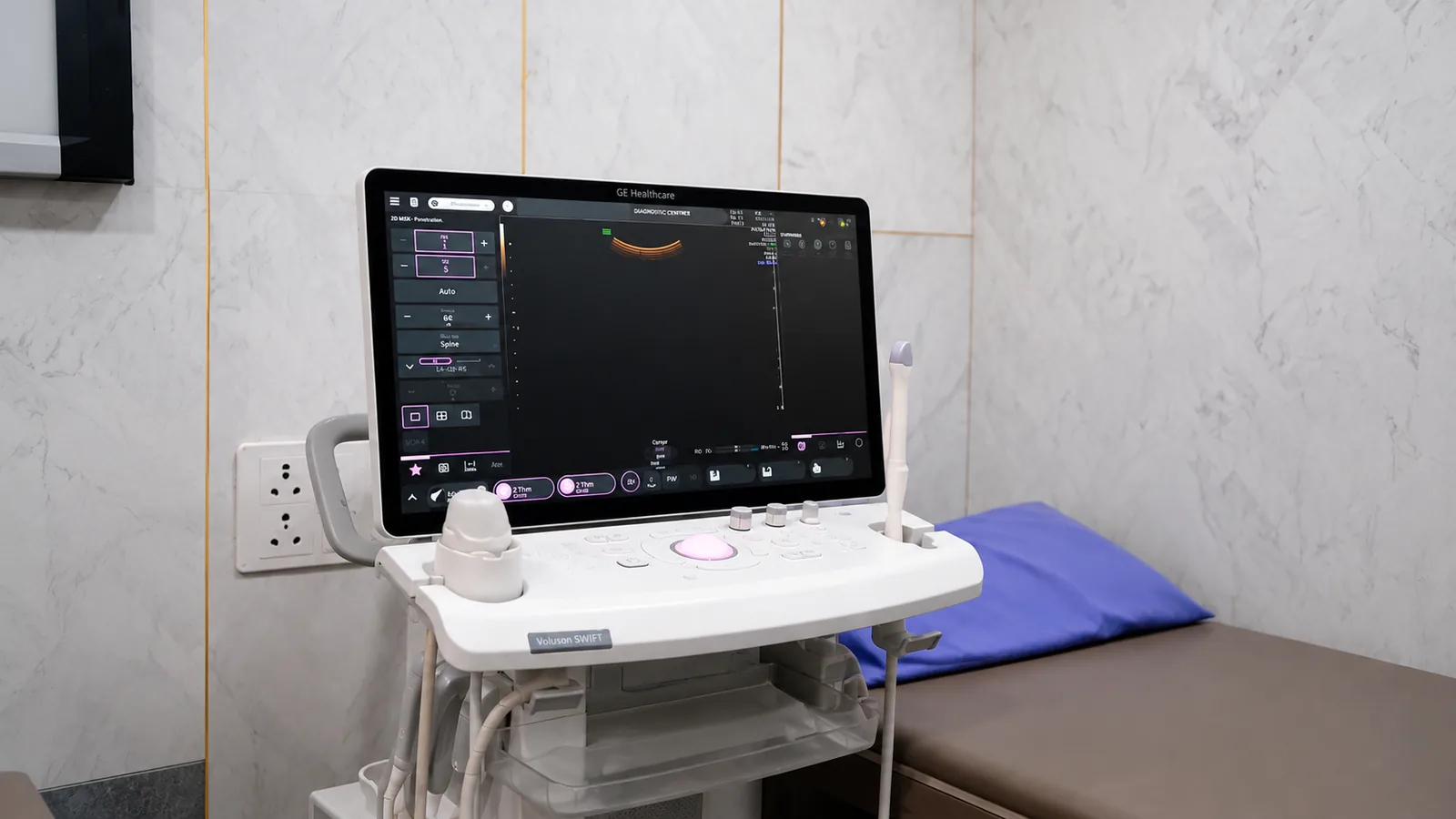
A standard ultrasound images the liver, gallbladder, kidneys, uterus, ovaries and other soft organs, and is the mainstay of pregnancy scanning from the early weeks through the anomaly scan.
Colour Doppler adds a map of blood flow on top of the image — showing whether an artery is narrowed, a vein is clotted, or blood is reaching an organ as it should. It turns a still picture into a study of movement.
Your doctor may recommend ultrasound or Doppler for
- Abdominal pain, gallstones, or liver and kidney assessment
- Pregnancy — dating, growth, and the anomaly scan
- Pelvic symptoms — uterus and ovaries
- A thyroid or breast lump, or other small-parts imaging
- Leg swelling or suspected clot, and arterial narrowing (Doppler)
How to prepare
Preparation depends on the scan. When you book, we will tell you exactly what yours needs — the two most common are below.
- Upper abdomen (liver, gallbladder): fast for 6–8 hours so the gallbladder is full
- Lower abdomen / pelvis and early pregnancy: come with a full bladder — drink water and do not pass urine beforehand
- Thyroid, breast, and Doppler studies: no special preparation
- Wear two-piece clothing so the area can be reached easily
- Bring previous scans for comparison
What to expect on the day
- 01
Settling in
You lie on the couch and the area is exposed. A water-based gel is applied so the probe makes good contact — it is cool but harmless.
- 02
The scan
The sonographer moves the probe over the skin, watching the live image. You may be asked to breathe in and hold, or turn onto your side.
- 03
Doppler, if included
For flow studies you may hear a rhythmic sound — that is blood flow being measured, and it is a normal part of the scan.
- 04
Afterwards
The gel is wiped off and you can dress and leave straight away. The radiologist's report follows.
Common questions
Is ultrasound safe in pregnancy?
Yes. Ultrasound uses sound waves, not radiation, and has been used safely in pregnancy for decades. It is the standard way to check a baby's growth and wellbeing.
Why do I need a full bladder for some scans?
For lower-abdomen and early-pregnancy scans, a full bladder pushes the bowel out of the way and creates a clear window to the pelvic organs, so the images are much better.
Does the scan hurt?
No. You feel the gentle pressure of the probe and the cool gel, nothing more. Some Doppler studies press a little more firmly to find a vessel, but it is not painful.
What is colour Doppler?
It is an ultrasound that also shows blood flow in colour — used to check for narrowed arteries, clots in veins, and whether blood is reaching an organ properly.
From the health library
Plain-language guides written by the team here — what the test measures, how to prepare, and what the result means.
 Pregnancy tests and scans in Lucknow: the antenatal schedule, explainedA long list of tests arrives with the first appointment and rarely with an explanation. Here is what each one is for.7 min read
 PCOS and hormone testing in Lucknow: which tests, and when in your cycleTiming changes the result. Which hormone tests matter for PCOS, which day to take them, and what the diagnosis actually requires.6 min read
 Liver function test in Lucknow: fatty liver, and reading an LFT reportRaised enzymes on a routine panel are common and rarely explained properly. Here is what each line means, and what usually causes it.6 min read