
Renal Scan in Lucknow — DTPA & DMSA Kidney Imaging
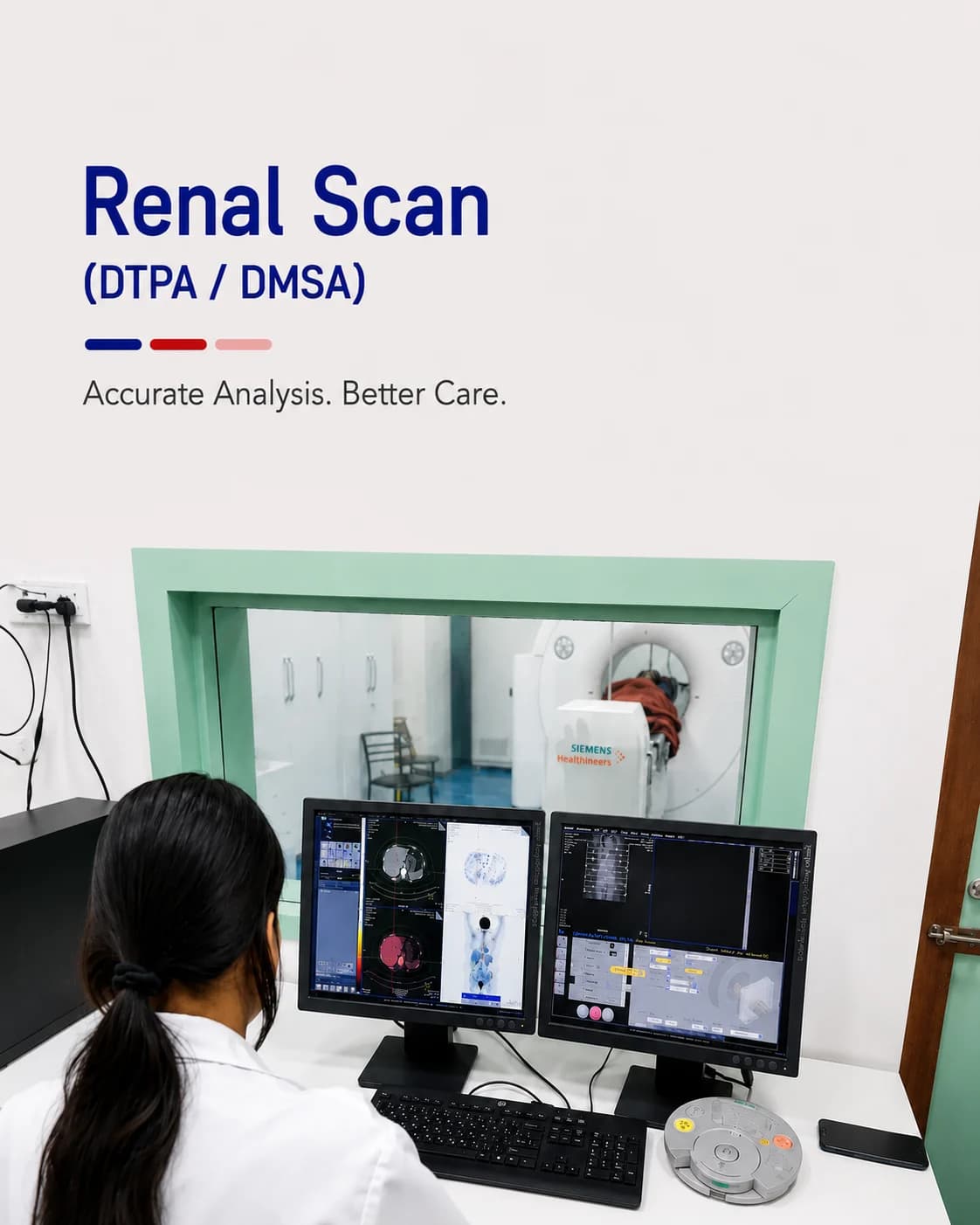
Measuring how well each kidney drains and works — one side against the other.
A nuclear renal scan measures how well each kidney is actually working and draining — separately, so the function of the left can be compared with the right. A blood test gives one combined number for both kidneys; this scan tells them apart, which is often exactly what a doctor needs to know.
At Charak, renal scans use a DTPA tracer to study drainage and filtration, or a DMSA tracer to look for scarring, and are reported by nuclear medicine physicians. They are especially useful in children with recurrent urine infection and in anyone with a suspected blockage.
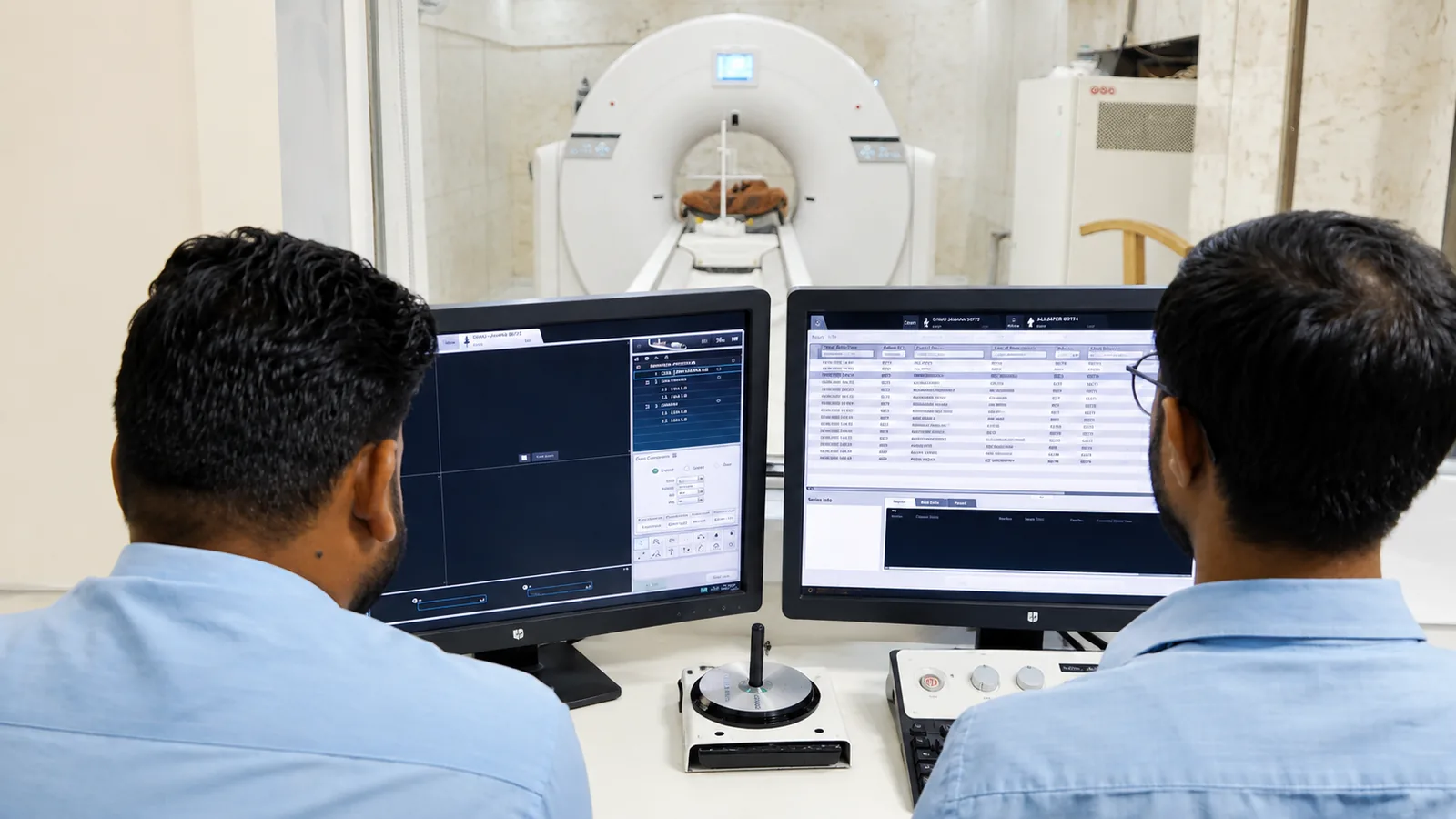
DTPA and DMSA — two questions, two tracers
A DTPA (dynamic) study follows the tracer as it is filtered and drains away, showing the filtration rate of each kidney and whether the outflow is obstructed — it captures the kidney in motion.
A DMSA (static) study shows the working kidney tissue itself, mapping any scarring, particularly after infection. Between them they answer the two key questions: how well is each kidney working, and is it damaged or blocked?
A renal scan may be recommended for
- Comparing the function of one kidney against the other (split function)
- A suspected blockage to kidney drainage (obstruction)
- Recurrent urine infection in children — checking for scarring
- Assessing a kidney before or after surgery
- Evaluating high blood pressure of possible kidney origin
How to prepare
Being well hydrated is important for a renal scan — it helps the tracer move through the kidneys as it should. We give you clear instructions for your specific study when you book.
- Drink water and stay well hydrated before the scan, unless told otherwise
- You may be asked to empty your bladder just before imaging
- Continue your usual medicines unless your doctor advises a change
- Tell us if you are, or might be, pregnant, or are breastfeeding
What to expect on the day
- 01
Tracer injection
The appropriate tracer — DTPA or DMSA — is given through a vein in the arm.
- 02
Imaging
For a DTPA study, imaging begins straight away and follows the tracer in real time; a DMSA study images after a delay.
- 03
Diuretic, if needed
In a drainage study a diuretic may be given during the scan to see how the kidney empties under a challenge.
- 04
Afterwards
You can resume normal activity; drinking water helps clear the small amount of tracer.
Common questions
How is a renal scan better than a blood test for the kidneys?
A blood test (like creatinine) gives one combined figure for both kidneys. A renal scan measures each kidney separately, so it can show that one is doing most of the work while the other is failing — which a blood test cannot reveal.
What is the difference between DTPA and DMSA?
A DTPA study is dynamic — it watches the tracer being filtered and draining, and detects obstruction. A DMSA study is static — it maps the working kidney tissue and shows scarring. Your doctor chooses based on the question being asked.
Why is a diuretic sometimes given during the scan?
In a drainage study, a diuretic makes the kidney work harder, which helps distinguish a true blockage from a kidney that is simply slow to empty — an important distinction for treatment.
From the health library
Plain-language guides written by the team here — what the test measures, how to prepare, and what the result means.
Related services
Gamma Camera / SPECT
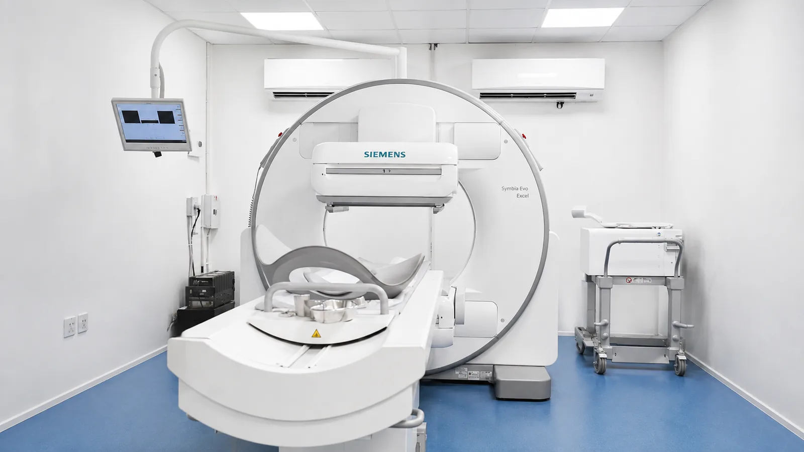
Functional imaging of the heart, bones, kidneys and thyroid using a gamma camera.
Read more →Bone Scan
A whole-skeleton study that finds active bone disease before an X-ray can.
Read more →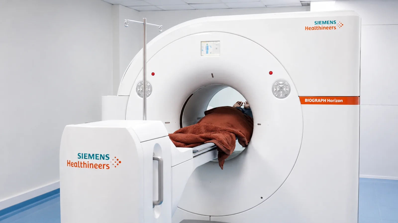
PET-CT Scan
Whole-body imaging that shows how tissue is functioning, not just how it looks.
Read more →