Nuclear Medicine
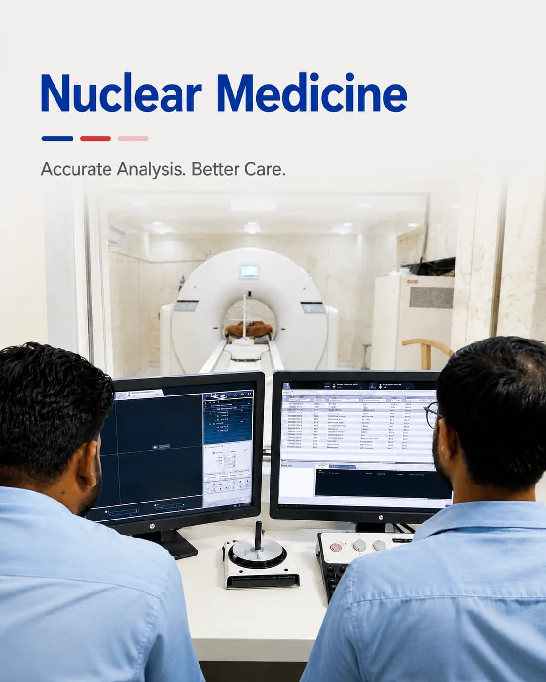
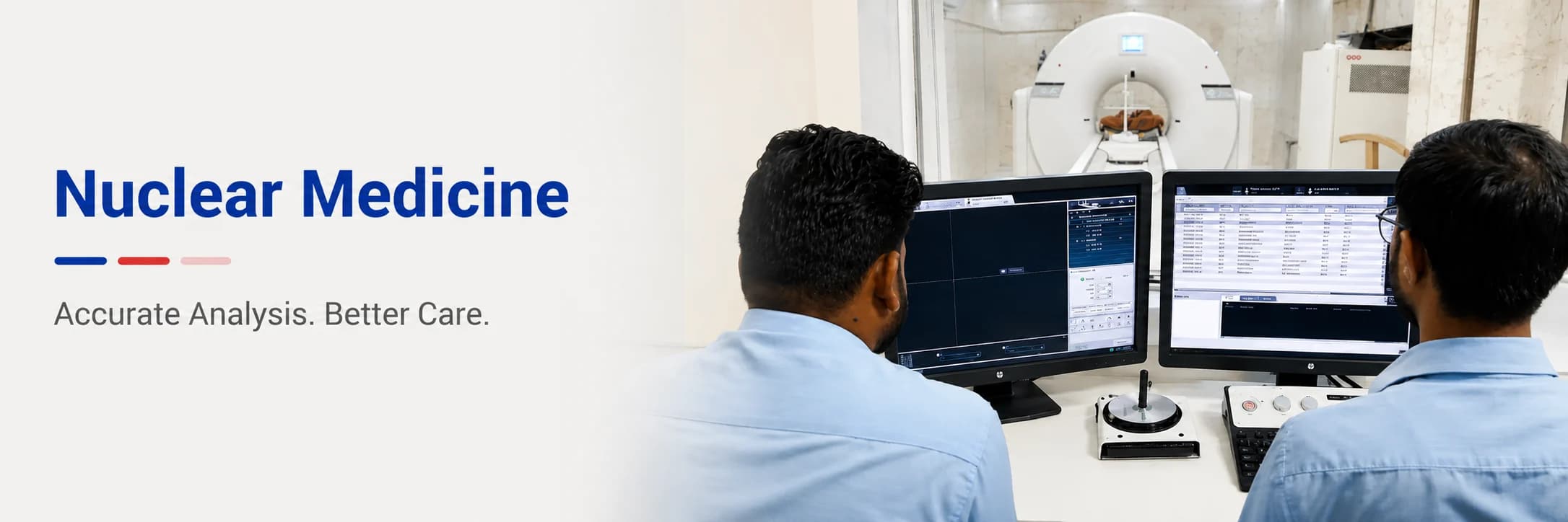
PET-CT and gamma camera studies that show how an organ is working, not only what it looks like — performed under strict radiation-safety protocol.
Functional imaging
Imaging that answers a different question
A CT or MRI shows structure. Nuclear medicine shows function — whether tissue is metabolically active, whether a kidney is draining, whether bone is remodelling. The two are complementary, and read together they answer questions neither can settle alone.
Function, not just anatomy
Tracers reveal metabolic activity, so disease can be detected before it changes an organ's shape.
Dose calculated per patient
Activity is matched to body weight and to the study, and recorded — the same discipline applied in CT.
Read with your other results
Scans are interpreted alongside the laboratory and imaging already on file at the centre.
Nuclear medicine
Explore each scan in detail
What each one involves, how to prepare, and what to expect on the day — on a dedicated page for every service.
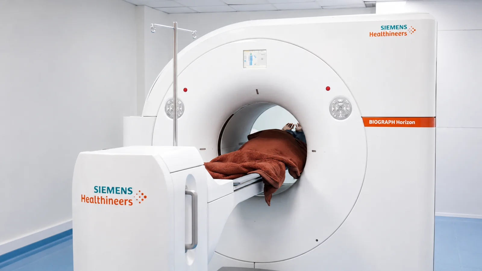
PET-CT Scan
Whole-body imaging that shows how tissue is functioning, not just how it looks.
Read more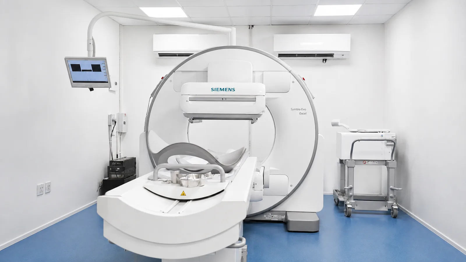
Gamma Camera / SPECT
Functional imaging of the heart, bones, kidneys and thyroid using a gamma camera.
Read more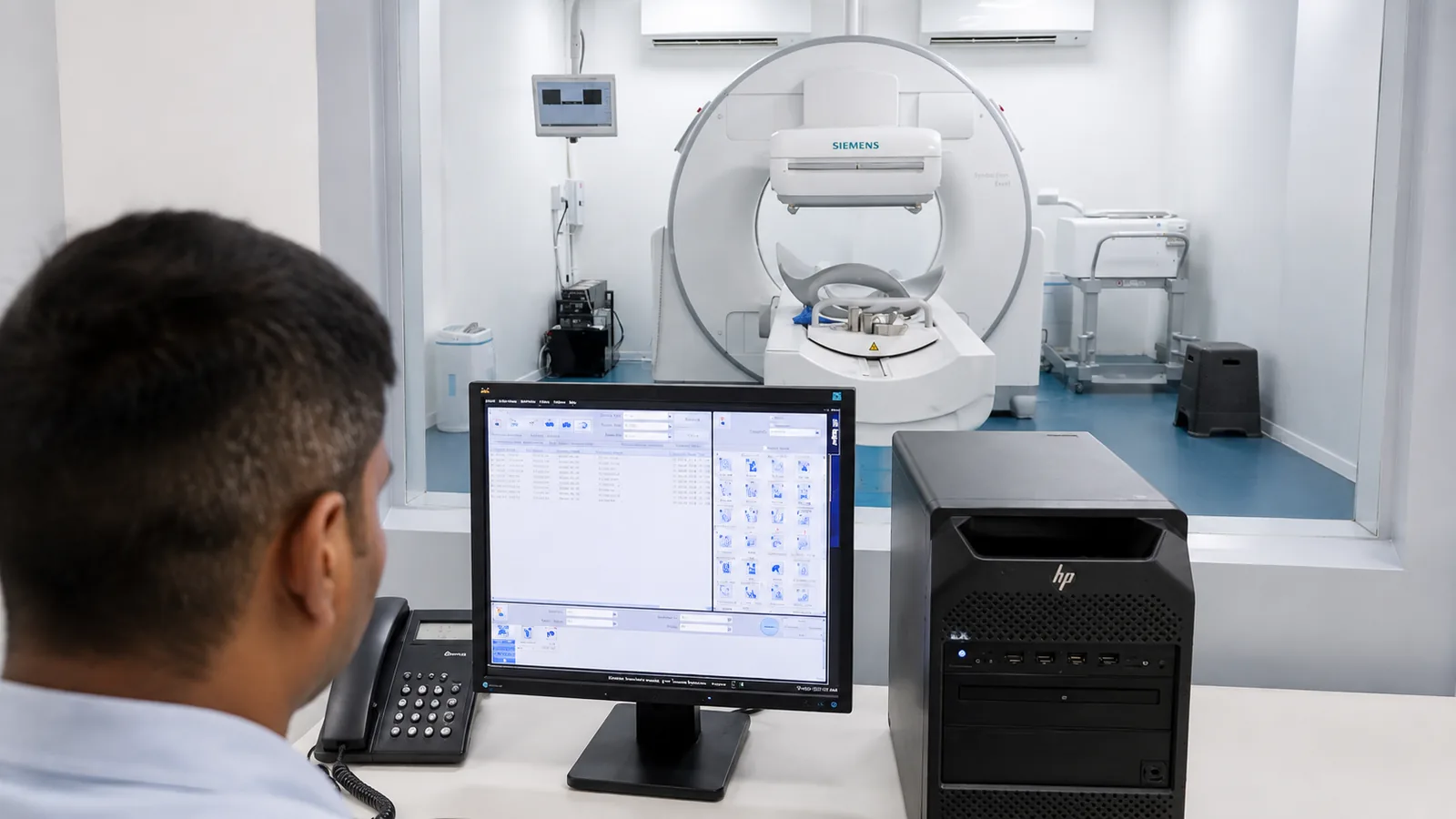
Thyroid Scan
A functional look at the thyroid gland — how active it is, and how it works.
Read moreBone Scan
A whole-skeleton study that finds active bone disease before an X-ray can.
Read more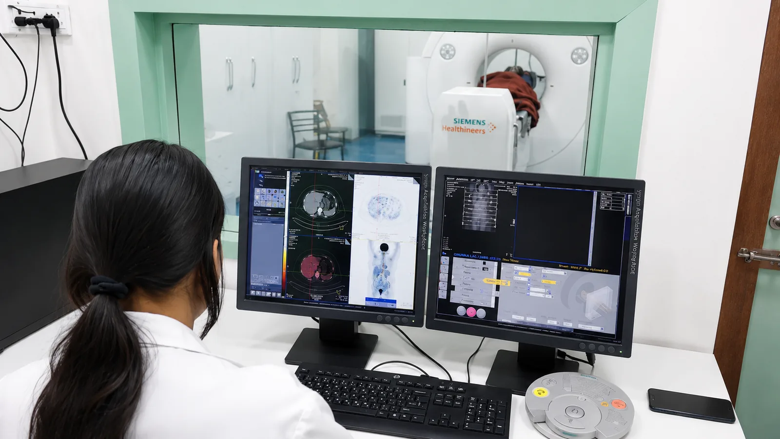
Renal Scan (DTPA / DMSA)
Measuring how well each kidney drains and works — one side against the other.
Read more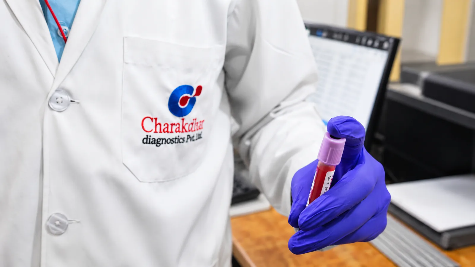
Radiation safety
What we do to keep the dose right
- The radiopharmaceutical dose is calculated for your body weight and the specific study — never a fixed default.
- Tracers are short-lived by design; activity falls away over hours, not weeks.
- Tell us if you are pregnant, might be pregnant, or are breastfeeding — some studies must be deferred or modified.
- Most studies involve a waiting period between injection and imaging; allow more time than a plain scan.
- You will be told what to drink, and what to avoid, both before the study and immediately after it.
If you are pregnant, think you may be pregnant, or are breastfeeding, tell the centre before your appointment rather than on the day. Some studies can be rescheduled, some can be modified, and a few should not be done at all — that decision needs to be made before a tracer is drawn up.